Arteriovenous malformation (AVM)
Find a neuro specialistArteriovenous malformation, or AVM, is an irregular connection between arteries and veins. Picture an AVM as tangled wires behind your television. The entanglement of these arteries and veins creates direct connections.
The direct connection can cause various problems if you have an AVM rupture, including a stroke. A stroke is a life-threatening medical emergency. Call 911 if you or someone you know is experiencing symptoms of a stroke.
What is an arteriovenous malformation?

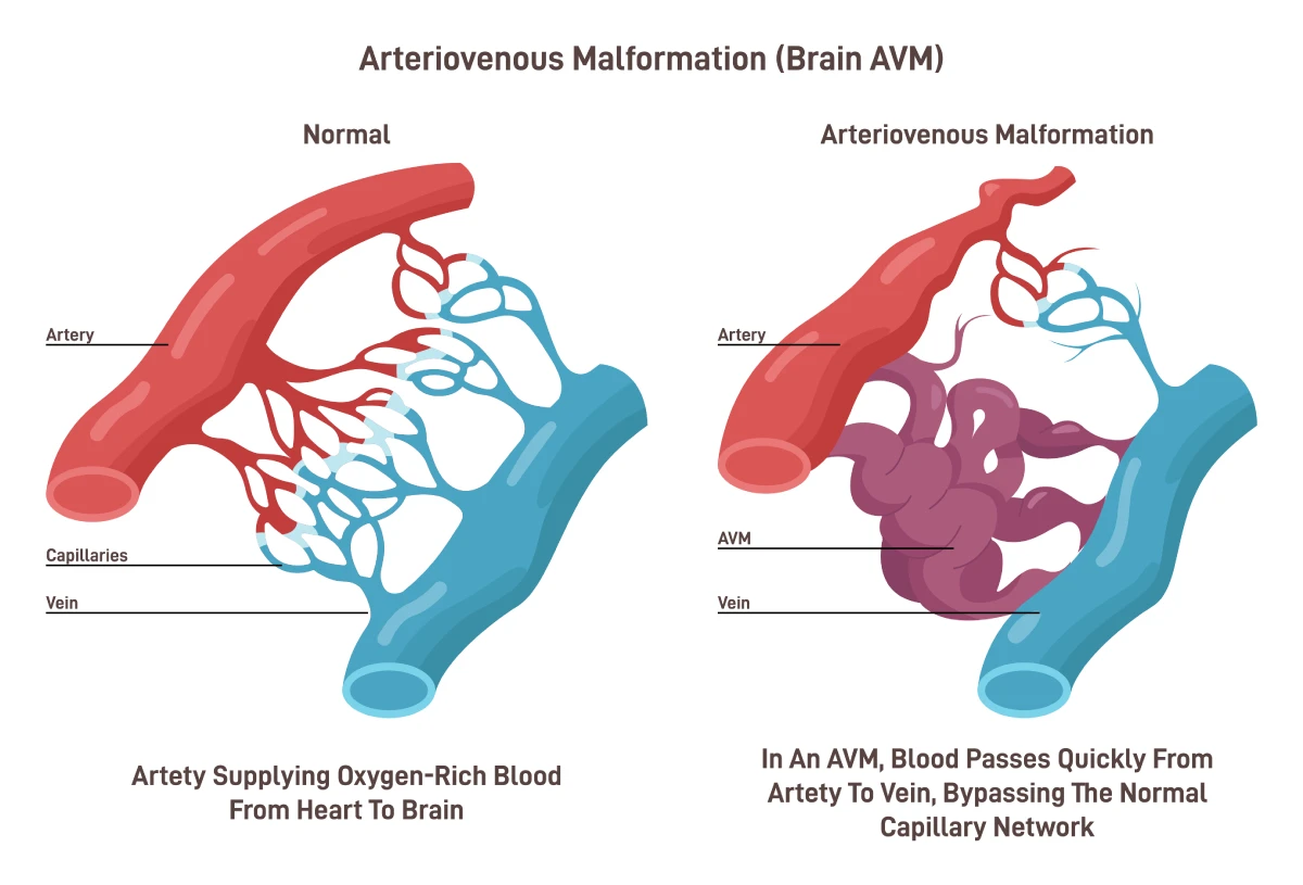
The image shows a normal connection between an artery and a vein compared to an arteriovenous malformation.
Your arteries carry blood from your heart. As the arteries move farther away from the heart, they branch off and get smaller. Very small arteries turn into capillaries that carry blood to your cells and tissues.
The capillaries have very thin walls and deliver nutrients and oxygen to your cells. Waste is brought into the capillaries and is sent back to the heart through larger veins.
An AVM is a tangle of blood vessels that bypasses the capillaries and connects the arteries directly to the veins. AVMs are common in the brain or spinal cord but can occur anywhere.
In AVM, this normal flow is disrupted, leading to a tangle of vessels that can cause various problems. An AVM can damage brain or spinal cord tissues through a brain hemorrhage, compression of the brain and spinal cord or reduced blood flow.
AVMs are more common in men, but anyone can be born with one.
Arteriovenous malformation symptoms
AVM symptoms vary based on their location and size. Some people do not have any symptoms, especially at an early age. People are usually in their mid-30s when symptoms first appear.
Symptoms may also appear or become worse during a pregnancy. This occurs due to pregnancy-related increases in blood volume and blood pressure.
Common AVM symptoms include:
- Confusion, hallucinations or dementia
- Headache
- Problems with memory, vision or speech
- Seizures
- Weakness or paralysis on one side of the body
For many people with an AVM, a hemorrhage is their first sign. If the bleeding becomes severe, it can cause a stroke.
Arteriovenous malformation complications
The greatest risk from an AVM is severe bleeding that leads to a stroke. However, an AVM can lead to other potential complications, such as:
- Bleeding in the brain
- Reduced oxygen to the brain
- Thin or weak blood vessels
- Brain damage
Diagnosing an arteriovenous malformation
To get an AVM diagnosis, your doctor will perform a physical exam and discuss any symptoms you’re having. They will review your medical history and listen to your heart. Some AVMs cause a noise your doctor may hear through a stethoscope.
Your provider may order image tests such as a CT scan or MRI to get detailed images of blood vessels in your spine, neck and head.
More advanced testing options include a cerebral angiogram. This test involves your doctor inserting a catheter in your neck or head to release a dye that highlights any malformations.
Arteriovenous malformation treatment
An AVM can be treated in several ways. If the AVM has not ruptured, your doctor will help you weigh the risks of possible hemorrhage against the risks of treatment.
Some factors that could increase your risk of a hemorrhage include:
- Pregnancy
- The location and size of the AVM
- Symptoms you are experiencing
- Your age and overall health
Your doctor may prescribe medications to help reduce symptoms, such as seizures and headaches.
In some cases, surgery is needed to repair an AVM. Surgical options include:
- Endovascular embolization: This is a minimally invasive arteriovenous malformation medical procedure where your doctor inserts a catheter into a blood vessel and guides it to the AVM. Your doctor injects glue or another material to plug the malformation. Typically, this procedure occurs before AVM removal through microsurgery.
- Microsurgery: Microsurgery involves removing a portion of the skull and using a surgical microscope and specialized tools to remove the AVM.
- CyberKnife® radiosurgery: This technique uses beams of radiation instead of open surgery to treat small, unruptured AVMs. Over a few months, the blood vessels deteriorate and close.
Following treatment, particularly in cases of a ruptured AVM, rehabilitation may be essential to help patients regain lost function and recover from any neurological deficits resulting from the AVM or its treatment. This could include physical therapy, occupational therapy or speech therapy.
Get care
We help you live well. And we’re here for you in person and online.